


Products
Biochemistry & Immunology
ELISA Kits
General
SKU :
Quantity
หมวดหมู่ : 1. Chemical and Reagents , Biochemicals , ELISA Kits & Assay Kits , Servicebio ,
แบรนด์ : Servicebio
Share
Product Information
Product Name | Cat. No. | Spec. |
Cortisol ELISA Kit (Common Species) | GE0002-48T | 48T |
GE0002-96T | 96T |
Product Description
Cortisol is a steroid hormone secreted by the adrenal cortex, also known as hydrocortisone.Cortisol plays a very important role in maintaining the stability of physiological functions and regulating the metabolism of protein, fat and sugar, and also has a strong anti-inflammatory, anti-shock and anti-allergic effect, with obvious circadian rhythm.Cortisol is needed by the body to maintain normal physiological functions during stress, so it is also known as the stress hormone.Cortisol ELISA Kit quantitatively assesses Cortisol in vitro in serum, plasma, tissue homogenate, cell lysate, cell culture supernatant, or other related liquids through competitive ELISA. Cortisolelisakit can detect both natural and synthetic Cortisol.
Storage and Shipping Conditions
Ship with wet ice; Store at 4, valid for 6 months; Use within 4 weeks once opened.
Product Components
Component Number | Component | GEH0014-48T | GEH0014-96T |
GE0002-1 | Microplate | 48T | 96T |
GE0002-2 | Standard | 1 vial | 2 vials |
GE0002-3 | Detection Antibody | 40 μL | 80 μL |
G0030 | Enzyme-labeled Antibody | 60 μL | 120 μL |
G0024 | Diluent A | 30 mL | 30 mL * 2 |
G0025 | Diluent B | 12 mL | 12 mL |
G0026 | TMB Substrate | 6 mL | 11 mL |
G0027 | Stop Solution | 6 mL | 6 mL |
G0028 | 25x Wash Buffer | 30 mL | 30 mL |
G6077 | Plate Sealers | 4 pcs | 4 pcs |
Manual | 1pc | 1pc | |
Additional Materials Required
1. Microtiter plate reader capable of measurement at 450nm. The reference wavelength is 630 nm(Refer to the instruction manual supplied with the instrument to pre-warm).
2. Single or multichannel pipettes and tips, loading slots, centrifuge tubes.
3. Deionized or distilled water.
4. Dry filter or absorbent paper.
5. Vortex mixer, microplate oscillator.
Sample Collection and Storage Instructions
1. Serum: Collect samples to clot for 30 minutes at room temperature before centrifugation at 1000 x g for 15 minutes at 2-8°C. Collect the supernatant to carry out the assay, make aliquots and store at -20 to avoid repeated freez-thaw cycles.
2. Plasma: Collect plasma with EDTA, sodium citrate or heparin as anticoagulants, and centrifuge at 1000 x g for 15 minutes at 2-8°C within 30 minutes of collection, aspirate supernatant for testing, make aliquots and store at -20°C to avoid repeated freez-thaw cycles.
3. Tissue homogenates: The tissues should be rinsed in pre-cold PBS to remove excess blood thoroughly. Weigh and mince into small pieces in homogenizer on ice. The homogenate was homogenised by adding 10 times the weight of the tissue in PBS and sonicated until clarified. Centrifuge at 12,000 x g for 5 minutes, discard the precipitate and aspirate supernatant to test, make aliquots and store at -20°C to avoid repeated freez-thaw cycles.
4. Cell lysates
a) For adherent cells: Wash the cells with PBS 2-3 times and aspirate the residual liquid thoroughly. Aspirate cell lysate at a ratio of 250 μL lysate per well of cells in 6-well plates and flasks, and shake repeatedly to bring the lysate into full contact with the cells for 3-5 minutes. Scrape off the cells with a cell spatula and collect in a centrifuge tube. Centrifuge at 12,000 x g for 5 minutes, aspirate supernatant to test, make aliquots and store at -20°C to avoid repeated freez-thaw cycles.
b) For suspension cells: Collect cells by centrifugation and mix with cell lysate at a ratio of 250 μL of lysate per well of a 6-well plate and shaken. Ice bath for 30 minutes, repeatedly pipetting every 10 minutes to ensure complete cell lysis. Centrifuge at 12,000 x g for 5 minutes and aspirate the supernatant to assay, make aliquots and store the supernatant at -20°C to avoid repeated freez-thaw cycles.
5. Cell culture supernatant: Centrifuge at 300 x g for 10 minutes and aspirate the supernatant to assay, make aliquots and store the supernatant at -20°C to avoid repeated freez-thaw cycles.
Note
1. Centrifuge to remove precipitate if sample is cloudy, not suitable for grossly hemolyzed and lipemic samples.
2. All the above samples should be sealed and stored for no more than 1 week at 4°C, 1 month at -20°C and 2 months at -80°C.
3. Gradually equilibrate frozen samples to room temperature before beginning assay.
Reagent Preparation
1. Remove the kit and samples from the refrigerator 20 minutes in advance and equilibrate to room temperature. Remove the plates and reagents as required for the experiment and store the remaining reagents at 4.
2. 1x Wash Buffer: Pour 30 mL of the Wash Buffer Concentrate (25x) into a 1000 mL graduated cylinder. Bring to final volume of 750 mL with distilled or deionized water. Mix gently to avoid foaming. Transfer to a clean wash bottle and store at 2-25.
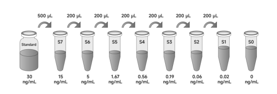
3. Preparation of standard product: Add diluent A to the standard product according to the volume marked on the label, and gently swirl to ensure full mixing.The standard product concentration after redissolution is 30ng/mL, which is the concentrated Cortisol standard product.Let stand for 10 minutes after remelting and mix well before diluting.
a) Preparation of standard curves for serum/plasma/tissue homogenate/cell lysate:
Fully mixed the redissolved standard product, 500µL concentrated Cortisol standard product was added to 500µL diluent A as the highest concentration S7 (15ng/mL) of the standard curve.Six 1.5mL centrifuge tubes (S1-S6) were arranged successively and 400µL diluent A was added to each tube.Draw 200µLS7 (15ng/mL) standard into the first centrifuge tube S6, gently blow and mix.Draw 200µL from S6 into the second centrifuge tube S5 and gently blow and mix.And so on to the standard product 3 times the ratio dilution.S0 is diluent A.
b) Preparation of standard curve of cell culture supernatant sample:
Fully mixed the redissolved standard product, 500µL concentrated standard product was added to 500µL cell medium as the highest concentration S7 (15ng/mL) on the standard curve.Six 1.5mL centrifuge tubes (S1-S6) were sequentially arranged and 400µL cell medium was added to each.Draw 200µLS7 (15ng/mL) standard into the first centrifuge tube S6, gently blow and mix.Draw 200µL from S6 into the second centrifuge tube S5 and gently blow and mix.And so on to the standard product 3 times the ratio dilution.S0 is the cell medium.
4. 1× antibody preparation: The antibody is briefly centrifuged and diluted to the working concentration by 1:100 times with diluent A. 1× antibody working liquid is mixed and prepared before clinical use.
5. Preparation of 1× enzymic antibody: The enzymic antibody is briefly centrifuged and diluted to the working concentration by 1:100 times with diluent B. 1× enzyme-labeled antibody working liquid is mixed and prepared before clinical use.
Note
1. Unopened kits: Store entirely at 4°C and valid for 12 months.
2. Opened kits: Lyophilised standards should not be reused after dissolution and remaining reagents should be stored promptly at 4°C and valid for 4 weeks. In addition, keep unused slats in an aluminium foil bag containing desiccant seal tightly and store at 4°C.
Assay Protocol
1. Add sample: Set up the standard wells, sample wells and blank wells separately. Dilute the standard product and sample with diluent A, set standard holes 7 holes (S1-S7), add 100μL standard product of different concentration to each hole successively, add 100μL diluent A to each blank hole, and add 100μL sample to each remaining hole.
2. Add antibody: Dilute antibody to working concentration with diluent A, add antibody 50μL per well.Sealing plate film sealing plate, 100-300rpm oscillation (ensure that each hole solution does not spill and can be fully mixed), temperature oscillation incubation for 1 hour.
Note: 1. Complete steps 1 and 2 as soon as possible with an interval of no more than 10 minutes. The sequence of steps cannot be changed.
2. Please refer to relevant literature to determine the approximate concentration of the protein to be detected in the sample. If the concentration is greater than or less than the maximum or minimum standard concentration of this kit, please carry out appropriate dilution or concentration before testing.
3. Wash plate: automatic washing plate or manual washing plate, each hole of the washing liquid is 300μL, injection and suction interval of 15-30 seconds.Wash the board five times.After the last plate washing is completed, the enzyme label plate is upside-down on the absorbent paper and patted dry properly, and the liquid in the hole is discarded;
4. Add the enzyme-labeled antibody: Dilute the enzyme-labeled antibody to the working concentration with diluant B, add the enzyme-labeled antibody working solution (prepared before use) 100μL in each well, replace the sealing plate with a new sealing plate, shake at 100-300rpm (ensure that the solution does not spill in each well and can be fully mixed), and incubate at room temperature for 30 minutes;
5. Plate washing: Repeat step 3.
6. Add TMB Substrate: Add TMB substrate solution 90μL to each hole, replace the new sealing plate film, and color rendering at room temperature (the reaction time should be controlled within 10-30 minutes, not more than 30 minutes.When the first 3-4 holes of the standard product have obvious gradient blue, the gradient of the last 3-4 holes is not obvious, it can be terminated);
7. Add Stop Solution: Add termination solution 50μL to each well to terminate the reaction, and the blue immediately turns to yellow.The addition sequence of the termination solution should be as close as possible to the addition sequence of the substrate solution.If the color is uneven, please gently shake the label plate to make the solution evenly mixed;
8. Reading: .After ensuring that there are no water droplets at the bottom of the enzyme label plate and no bubbles in the hole, the detection wavelength of 450nm is used to read the value within 10 minutes.It is recommended to use dual wavelengths, namely the detection wavelength of 450nm, the reference wavelength, or the correction wavelength of 630nm to read the value at the same time. Using only 450nm will reduce the accuracy.
Note
1. Store kits according to instructions. Gradually equilibrate samples to room temperature for 20-30 minutes before beginning assay.
2. The standards are lyophilised powders. The diluent volume and ratio should strictly follow the kit instructions. To ensure the accuracy of the standards, please do not reuse if they are left over.
3. The 25x Wash Buffer may crystallise at low temperatures. If crystals have formed in the Buffer Concentrates, warm them gently until they have completely dissolved before prepare to working solution.
4. TMB is Hazardous. Care should be taken to avoid contact with skin or eyes. In the case of contact with skin or eyes wash immediately with water.
5. Stop Solution contains acid, care should be taken to avoid contact with skin or eyes. In the case of contact with skin or eyes wash immediately with water.
6. An intense aqua blue color indicates that the TMB Sbustrate is contaminated, do not use it.
7. Reagents are lot-specific. Do not mix or interchange different reagent lots from various kit lots.
8. All reagents must be at room temperature (25-28). Temperature below 25will result in a significant decrease in the absorbance of the final detection.
9. Repeat wells are recommended for standards and samples to ensure confidence in the test results.
10. Do not expose kit reagents to strong light during storage or incubation.
11. Mix gently after sample addition to avoid foaming.
12. Wear suitable protective clothing such as laboratory overalls, safety glasses and gloves.
13. For research use only. Not for use in diagnostic or therapeutic procedures.
14. To avoid microbial contamination or cross-contamination of reagents or samples that may invalidate the test, use disposable pipette tips for each transfer.
15. After the last wash step, empty wells and tap microwell strips on absorbent pad or paper towel to remove excess Wash Buffers. Do not put the absorbent paper directly into the wells to absorb water.
Results Analysis
1. Results calculation
a) It is recommended to calculate the average absorbance values for each set of duplicate standards and samples to obtain more accurate results.
b) Create a standard curve with curve-fitting statistical software by plotting the standard concentration on the abscissa against the OD value on the ordinate. the closer the correlation coefficient R value is to 1, the better the fitting effect is. The best fitting curve was determined by regression analysis.
c) The sample concentration is calculated by substituting the OD value. If the test sample was diluted, multiply the appropriate dilution factor for actual concentration.
d) The standard curve can be linearised by taking a logarithmic fit to the concentration values and OD values. This process may result in more sample concentrations, but the accuracy of the data will be reduced.
2. Typical data
Perform a standard curve with each assay. The OD values of the standard curve may vary according to the conditions of assay performance (e.g., operator, pipetting technique, washing technique, or temperature effects). The standard curves provided in the instructions are for reference only.
ng/mL | OD | Average | |
15 | 0.070 | 0.064 | 0.067 |
5 | 0.159 | 0.143 | 0.151 |
1.67 | 0.383 | 0.371 | 0.377 |
0.56 | 0.789 | 0.795 | 0.792 |
0.19 | 1.364 | 1.368 | 1.366 |
0.06 | 1.709 | 1.681 | 1.695 |
0.02 | 1.923 | 1.986 | 1.955 |
0 | 2.219 | 2.203 | 2.211 |

3. Sensitivity
The lowest detectable concentration of Cortisol is 8.6pg/mL(the average of three independent experiments).This value is the average OD value measured by 20 blank holes plus the concentration value corresponding to twice SD
4. Precision
Precision is expressed as the coefficient of variation(CV) of the measured values of the samples. CV(%)=(SD/Mean)×100.
Intra-assay Precision: 3 samples with low, mid and high level were assayed 20 times in multiple assays to determine precision between assays. Intra-assay Precision: CV<9%.
Inter-assay Precision: 3 samples with low, mid and high level were assayed in replicates of 8 to determine precision within an assay. Inter-assay Precision: CV<15%.
5. Spike recovery
A certain amount of Cortisol (labeled samples) was added to 5 sets of normal human serum, and the mean value was measured repeatedly, and the recovery rate was calculated (the ratio of measured value to theoretical value) for the serum without Cortisol.Recovery rates range from 80% to 107%, with an average recovery rate of 92%.
6. Linearity of dilution
A certain amount of Cortisol (labeled samples) was added to five sets of human serum and diluted in a series within the kinetic range of a standard curve to evaluate the linearity of the test.The linear range is the ratio of Cortisol content measured to theoretical value in a diluted sample.
Dilution | Mean(%) | Range(%) |
1:2 | 93 | 85-108 |
1:4 | 95 | 89-101 |
1:8 | 108 | 94-118 |
1:16 | 105 | 99-115 |
7. Sample values
The serum/plasma samples of 30 healthy volunteers were analyzed with this kit. The drug use history of the volunteers was unknown, and all samples were positive.
8. Specificity
This kit is used to detect Cortisol, which can identify natural and synthetic Cortisol, and has no obvious cross-reaction with Progesterone, Testosterone, Estradiol, etc.Due to technical and sample source limitations, it is not possible to perform cross-reaction tests for all related or similar substances, so this kit may cross-react with other substances that have not been tested.
Test Protocol Summary
1. Prepare standards, reagents and samples according to instructions before the experiment.
2. Add sample (standard product or sample) 100μL/ well, then add antibody working solution 50μL/ well, and add sample continuously within 10 minutes.After mixing, shake at room temperature for 1 hour;
3. After washing for 5 times, pat dry, add enzyme-labeled antibody working solution 100μL/ well, and shake at room temperature for 30 minutes;
4. Wash for 5 times and pat dry, add TMB substrate 90μL/ well, incubate at room temperature for 10-30 minutes away from light;
5. Add termination solution 50μL/ well;
6. The OD value is detected at 450 nm wavelength within 10 minutes, with a reference wavelength of 630 nm.
Layout
1 | 2 | 3 | 4 | 5 | 6 | 7 | 8 | 9 | 10 | 11 | 12 | |
A | S7 | S7 | ||||||||||
B | S6 | S6 | ||||||||||
C | S5 | S5 | ||||||||||
D | S4 | S4 | ||||||||||
E | S3 | S3 | ||||||||||
F | S2 | S2 | ||||||||||
G | S1 | S1 | ||||||||||
H | S0 | S0 |


